Top technologies improving breast cancer screening today combine better imaging, smarter software, and more personalised risk tools to find cancers earlier, especially in women with dense breasts or higher risk. Instead of replacing mammography, most of these innovations build on it, improving accuracy, reducing false positives, and helping radiologists focus on the most suspicious cases.
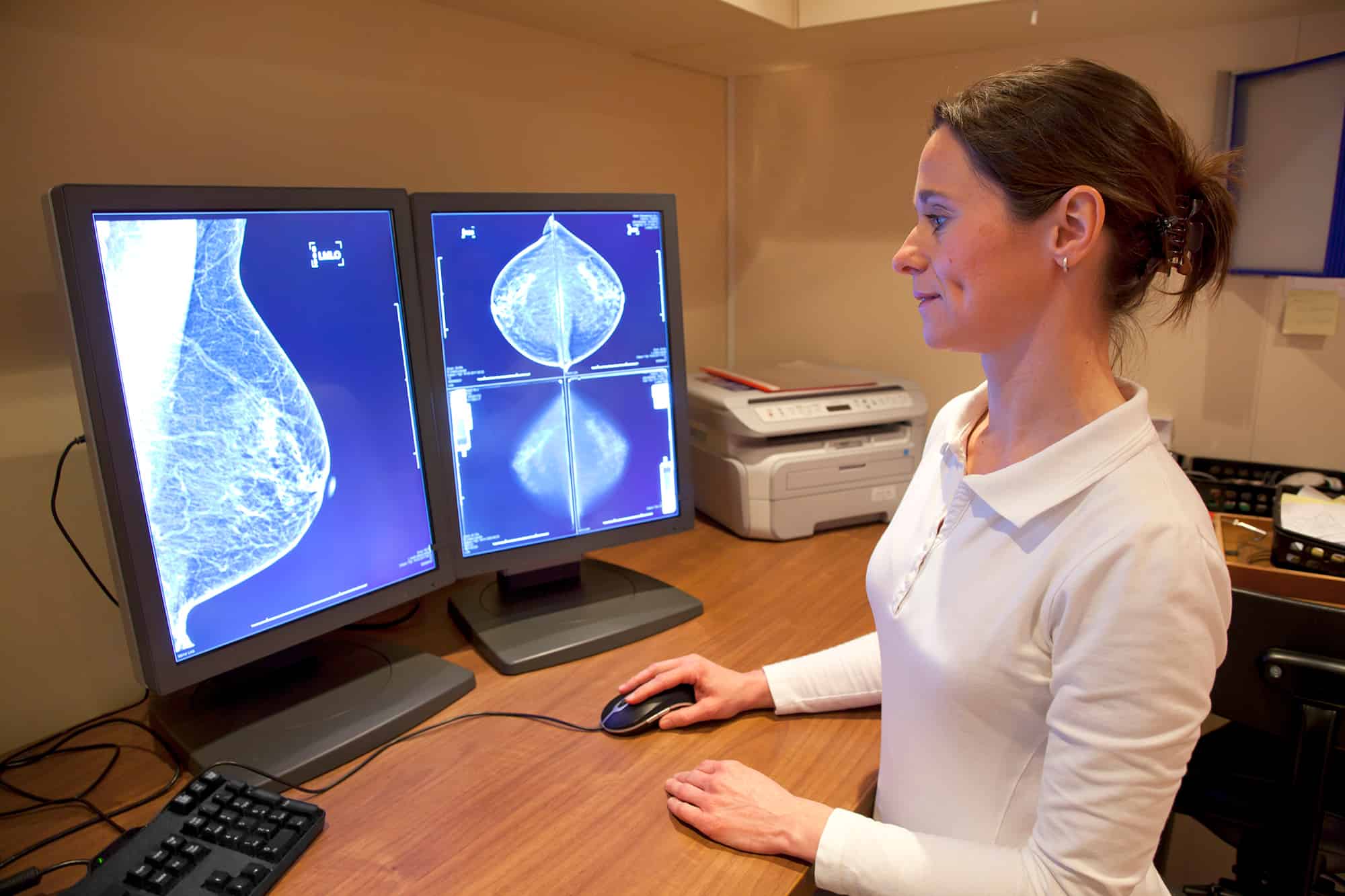
Digital and 3D mammography
Digital mammography is still the backbone of breast cancer screening worldwide, but the technology has become far more advanced than older film systems. Modern full‑field digital mammography delivers clearer images, better contrast and more flexible image processing, which helps radiologists detect small tumors and subtle distortions earlier.
A major upgrade is 3D mammography, also called digital breast tomosynthesis (DBT). DBT acquires multiple low‑dose X‑ray images from different angles and reconstructs them into thin “slices” of the breast, reducing the problem of overlapping tissue that can hide cancers in standard 2D views.
Large trials and meta‑analyses show that DBT improves sensitivity and cancer detection rates compared with digital mammography alone, while also lowering false‑positive recalls. Clinics report that 3D mammography is especially helpful in women with dense breasts, where conventional 2D images are harder to read.
AI‑assisted mammography
Artificial intelligence is one of the most important changes in breast imaging in the last few years. Deep‑learning systems can analyse mammograms and tomosynthesis images, highlight suspicious areas, and provide decision support scores that help radiologists prioritise and scrutinise the right regions.
Reader studies and early clinical trials indicate that AI support can increase cancer detection rates and reduce abnormal interpretation rates when used alongside radiologists. The MASAI randomised trial in the Swedish national screening programme, for example, reported a 29% increase in cancer detection and a 44% reduction in radiologist reading workload when an AI system (Transpara) was integrated into the workflow.
AI is also being used for risk‑stratified screening. In February 2026, the Clairity model became the first FDA‑authorised AI platform to estimate a woman’s five‑year breast cancer risk directly from a screening mammogram, enabling personalised screening intervals and supplemental imaging decisions. These tools move screening away from a one‑size‑fits‑all model and toward tailored surveillance based on individual risk.
Automated breast ultrasound (ABUS)
Dense breast tissue can mask tumours on mammograms because both appear white on X‑ray images. Automated breast ultrasound (ABUS) was developed as a supplemental screening technology to address this gap, particularly for women with dense breasts and average to intermediate risk.
ABUS uses a large, automated transducer to capture 3D ultrasound volumes of the entire breast, standardising the exam and producing datasets that radiologists can review slice by slice. GE’s Invenia ABUS 2.0 is the first FDA‑approved ABUS system for supplemental screening in women with dense breasts, and clinical studies show that adding ABUS to mammography can improve cancer detection by more than 35% compared with mammography alone.
Because ultrasound uses sound waves rather than ionising radiation and does not require compression in the same way as mammography, ABUS can be more comfortable for some patients and is particularly useful in identifying small invasive cancers hidden in dense tissue.
Advanced MRI and contrast‑enhanced techniques
Magnetic resonance imaging (MRI) is a highly sensitive tool for breast cancer detection, especially in high‑risk women, though it is not used as a universal screening test due to cost and resource demands. Modern breast MRI techniques with dynamic contrast enhancement and diffusion‑weighted imaging can identify cancers that may be missed on mammography and ultrasound, particularly in dense breasts and in younger women.
At the same time, contrast‑enhanced mammography (CEM) is emerging as a more accessible alternative in some centres. CEM uses iodine contrast with digital mammography to show areas of increased blood flow that may correspond to tumours, offering some of the functional information of MRI with mammography‑like workflows and equipment. These contrast‑based methods improve lesion conspicuity and can help clarify equivocal findings on standard imaging.
New sensor‑based and microwave imaging
Beyond established modalities, researchers are developing sensor‑based and microwave imaging systems to make screening more comfortable and accessible. An example is the MammoWave / MammoScreen project, which uses non‑ionising microwave signals to image breast tissue without compression, with the goal of reaching sensitivity above 90% and specificity above 95% in large population‑based trials.
A recent review in Frontiers in Oncology highlights a broad pipeline of biosensors, optical sensors, and other non‑invasive technologies for low‑cost, accessible breast cancer screening, especially in low‑ and middle‑income settings. These devices aim to detect physiological changes such as temperature, vascular patterns, or electrical properties that may indicate early tumour growth, offering an alternative or complement to conventional imaging.
Better workflows and access technologies
Technology is also improving how screening is delivered, not just how images are captured. Digital platforms, structured reporting tools, and integrated PACS/RIS systems are making it easier to track results, standardise BI‑RADS assessments, and ensure follow‑up of abnormal findings.
Mobile mammography units equipped with modern digital and 3D systems are expanding access in rural and underserved areas, making it more likely that women will receive routine screening on schedule. Some programmes now combine mobile units with tele‑radiology and AI triage so that images can be interpreted quickly even where specialist breast radiologists are scarce.
At the same time, risk‑assessment software that integrates family history, genetics, prior imaging and lifestyle factors is being used to identify women who might benefit from earlier or more intensive screening schedules. This helps direct advanced technologies such as MRI or ABUS to those most likely to benefit, improving both efficiency and outcomes.
How these technologies fit together
Taken together, today’s top screening technologies are not about replacing mammography with a single new modality, but about combining tools intelligently:
- Digital and 3D mammography remain the primary population‑level screening methods.
- AI systems enhance mammography and DBT by improving detection, reducing workload, and enabling personalised risk prediction.
- ABUS and ultrasound supplement mammography in women with dense breasts to uncover cancers masked on X‑ray.
- MRI and contrast‑enhanced techniques provide high‑sensitivity options for high‑risk women or complex cases.
- Emerging sensor‑based and microwave imaging aim to make screening more comfortable and accessible.
The end result is a screening ecosystem that is more accurate, more personalised and more inclusive than in the past. By catching cancers earlier and reducing unnecessary recalls, these technologies are already helping improve breast cancer outcomes and are likely to play an even larger role in the years ahead.